Volume: 16 Issue: 4 August 2018
FULL TEXT
Objectives: De novo steatosis after liver transplant is common and can occur in up to one-third of patients who are transplanted for liver disease other than for nonalcoholic fatty liver disease. Genetic factors may influence posttransplant steatosis; in a posttransplant setting, donor or recipient genetic factors could also play roles. Genetic polymorphisms in the adiponectin gene have been associated with metabolic syndrome in the pretransplant setting. We aimed to assess the association between donor and recipient adiponectin polymorphisms and early posttransplant hepatic steatosis identified on liver biopsies.
Materials and Methods: Clinical data were collected for 302 liver transplant patients who underwent protocol biopsies for hepatitis C. Of these, 111 patients had available biopsies and donor/recipient DNA. Patients with grade 1 steatosis or greater (35% of patients) were compared with patients without posttransplant steatosis with respect to clinical features and donor/recipient adiponectin polymorphism genotypes.
Results: Patients who developed posttransplant steatosis and those without steatosis were similar with respect to individual components of metabolic syndrome. The adiponectin polymorphisms rs1501299 G/G and rs17300539 G/G genotypes in recipients were associated with early posttransplant graft steatosis. We found no associations between graft steatosis and donor adiponectin polymorphisms.
Conclusions: Genetic polymorphisms in the adiponectin gene of recipients (but not donors) are associated with early de novo posttransplant hepatic steatosis, independent of components of metabolic syndrome.
Key words : Graft steatosis, Hepatitis C, Liver transplantation
Introduction
Posttransplant nonalcoholic fatty liver disease (NAFLD) is a common complication after liver transplant.1 Nearly 1 of 3 patients transplanted for a non-NAFLD indication will develop fatty liver disease after transplant, which may predispose patients to fibrosis progression even when their original liver disease is under good control.2 Posttransplant metabolic syndrome and steatosis are precursors to steatohepatitis in the allograft.1 Graft steatosis is partly driven by posttransplant metabolic syndrome, which in turn is influenced by weight gain, drug-induced hyperlipidemia, and diabetes, often related to immunosuppression.1 However, the incidence of NAFLD is different among patients with similar body mass index and metabolic risk factors, raising the possibility of genetic factors driving posttransplant NAFLD mediated both by metabolic syndrome and independent of it.
Liver biopsy remains the criterion standard for assessing disease recurrence and severity after liver transplant; in the absence of reliable noninvasive biomarkers of disease severity, many transplant programs have adopted protocol liver biopsy to risk-stratify patients before the availability of effective direct-acting antiviral therapies.3 These biopsies, performed routinely in all patients with hepatitis C virus (HCV) regardless of liver enzymes, allow identification of de novo hepatic steatosis in the posttransplant setting. Plasma adiponectin levels have been associated with different genetic loci, but the adiponectin gene (ADIPOQ), located at position 3q27, remains the main genetic determinant of plasma levels.4,5 Several single nucleotide polymorphisms located in the ADIPOQ gene have been associated with adiponectin serum levels and obesity, making this gene a candidate for obesity and metabolic syndrome.6
Genetic polymorphisms in the adiponectin gene have been hypothesized to be a risk factor for NAFLD. Our aim was to assess the relationship between donor and recipient genetic polymorphisms in the adiponectin gene and posttransplant hepatic steatosis. We hypothesized that posttransplant hepatic steatosis is driven by metabolic syndrome in the recipient rather than donor factors and therefore likely associated with recipient adiponectin genetic polymorphisms.
Materials and Methods
Study design
We reviewed 302 adult patients (> 18 years old) transplanted for cirrhosis
secondary to chronic hepatitis C between 2006 and 2011 at a single tertiary care
academic medical center (Cleveland Clinic, Cleveland, OH, USA). This was a
retrospective analysis of a prospectively assembled cohort. All patients with
chronic HCV who underwent protocol liver biopsies at 6 months and were at 1-year
posttransplant were identified. Patients who had a self-reported posttransplant
alcohol intake of > 30 g/week were excluded. Patients who lacked donor and
recipient DNA results or posttransplant protocol liver biopsies were also
excluded.
Liver biopsy
A single pathologist with expertise in posttransplant liver biopsies who was
blinded to the adiponectin polymorphism and clinical data interpreted all
biopsies. Hepatic steatosis was graded on an ordinal scale from 0 to 3 (grade 0
= less than 5% steatosis; grade 1 = 5%-33% steatosis; grade 2 = 34%-66%; and
grade 3 = greater than or equal to 67% steatosis).7 Hepatic fibrosis was
quantified from stages 0 to 4, with advanced fibrosis defined as fibrosis stage
≥ 3 and graft cirrhosis defined as stage 4.
Posttransplant immunosuppression mainly included tacrolimus, with mycophenolate added for 12 months. Prednisone was weaned within 2 months after transplant. Patients who received transplants for hepatocellular carcinoma and those with chronic kidney disease were considered high risk for recurrence and were switched to sirolimus after 12 weeks based on the discretion of the treating hepatologist.
The study protocol conformed to the ethical guidelines of the 1975 Declaration of Helsinki and 2008 Declaration of Istanbul and was approved by the Institutional Review Board at the Cleveland Clinic.
DNA collection and genotyping
Donor and recipient DNA results were extracted from whole blood and stored at
the time of transplant according to the center’s standard protocol. Adiponectin
gene polymorphisms rs1501299 (+276G>T), rs266729 (-11377C>G), rs2241766
(+45T>G), and rs17300539 (-11391 G>A) were analyzed by TaqMan single nucleotide
polymorphism genotyping assay (Life Technologies, Carlsbad, CA, USA).
Statistical analyses
Data are presented as mean and standard deviation, median (with 25th to 75th
percentiles), or number and percent (%). The prevalence of each genotype among
donors and recipients was estimated by calculating the percentage of patients
with each genotype. Pearson chi-square tests were used for categorical
variables, and analysis of variance or
the nonparametric Kruskal-Wallis tests were used
for continuous or ordinal factors. In addition, multivariable logistic
regression was performed to assess associations between genotypes and steatosis
after adjusting for race and HCV genotype. P < .05 was considered statistically
significant. All analyses were performed using SAS (version 9.4, The SAS
Institute, Cary, NC, USA).
Results
Recipient characteristics
For this study, we evaluated 302 consecutive patients who received transplants
for cirrhosis of the liver between 2006 and 2011. Of these, 111 donor/recipient
pairs who met the inclusion criteria were included in the analysis. Mean age of
recipients at time of transplant was 56 ± 6 years and 73% were male. There were
39 patients noted to have grade 1 or greater steatosis in protocol
posttransplant liver biopsies, whereas the other 72 patients did not have
posttransplant steatosis. Patients with posttransplant steatosis were more
likely to be white and less likely to have genotype 1 infection (Table 1). There
were no other significant differences between the groups, including coexisting
pretransplant liver disease (alcohol/nonalcoholic steatohepatitis [NASH]) or
the presence of risk factors for metabolic syndrome (Table 1).
Donor characteristics
Patients with and without steatosis were compared with respect to donor
characteristics (Table 2). Median donor age was 40.8 ± 14.0 years, and 75% of
all donors were white. There were no significant differences between the 2
groups. The median donor risk index was 2.1, which was comparable between the 2
groups.
Posttransplant immunosuppression and metabolic syndrome
There were no differences between the 2 groups in terms of immunosuppression
used. Both groups were comparable in terms of components of posttransplant
metabolic syndrome, including body mass index, diabetes, hypertension,
triglycerides, and high-density and low-density lipoprotein levels assessed at 1
year posttransplant (Table 3).
Recipient adiponectin polymorphisms
The adiponectin polymorphisms hypothesized to be associated with increased risk
of steatosis based on data in the nontransplant population included the
rs1501299 G/G, rs266729 non-C/C, rs17300539 G/G, and rs2241766 T/T
polymorphisms. On univariate analysis, the risk allele for 3 polymorphisms
(rs1501299, rs266729, rs17300539) in recipients was found to be associated with
early posttransplant steatosis (Table 4). The recipient rs1501299 G/G
polymorphism was more common among patients with posttransplant steatosis
(64.1%) than in those without (42.3%) (P = .028; Table 4). Similarly, the
recipient adiponectin rs266729 non-C/C genotype was significantly more common
among patients with steatosis (47% vs 22.5%; P = .008; Table 4). Finally, the
recipient rs17300539 G/G polymorphism was
more common among patients with steatosis (97.2%) than in patients without
steatosis (97.2% vs 81.9%; P = .026; Table 4). The recipient rs2241766 polymorphism did not differ between
the 2 groups.
On multivariate analysis, 2 recipient polymorphisms (rs1501299 and rs17300539) were found to be significantly associated with posttransplant steatosis (Table 5). After we adjusted for race and HCV genotype, we found that patients who developed posttransplant steatosis were 2.5 times more likely to have the rs1501299 G/G genotype than the non-G/G genotype (odds ratio = 2.5, 95% confidence interval, 1.02-6.2). Recipients with the rs17300539 polymorphism were 20.8 times more likely to have had the G/G genotype than the non-G/G genotype (odds ratio = 20.8, 95% confidence interval, 1.02-425.8). We found no association between any of the donor adiponectin polymorphisms and posttransplant steatosis.
Discussion
One-third of all patients transplanted for non-NASH indications develop de novo fatty liver disease after transplant. This is driven by metabolic syndrome but can also occur independent of it, with genetic factors likely contributing. Although genetic factors including adiponectin polymorphisms have been studied in nontransplant NAFLD, there are limited data in the posttransplant setting and particularly in the setting of patients transplanted for a non-NASH indication. Moreover, potential genetic contributions from the donor may also play a role in the posttransplant setting. Our data highlight the role of recipient adiponectin polymorphisms in the development of posttransplant hepatic steatosis, independent of individual components of metabolic syndrome.
The increased incidence among family members and high concordance rates among monozygotic twins with NAFLD indicate a genetic predisposition.8 The ADIPOQ gene is located on human 3q27 and leads to overexpression of adipocyte-specific secretory proteins.9 Multiple studies have demonstrated that lower adiponectin levels are associated with type 2 diabetes mellitus, dyslipidemia, and hypertension.10-12 Conversely, weight loss leads to an increase in plasma adiponectin levels.13 Treatment with peroxisome proliferator-activated receptor gamma 2 agonists for hyperglycemia in patients with type 2 diabetes mellitus and blockade of the renin-angiotensin system can increase plasma adiponectin levels.14,15 In the liver, adiponectin has been postulated to play hepatoprotective and anti-fibrotic roles, possibly through modulation of tumor necrosis factor α, nuclear factor κB, interleukin 6, and hepatic stellate cells.16-19
Previous cardiovascular studies in the literature have suggested an association between adiponectin genetic polymorphisms and metabolic syndrome.20 Although our study did not explore the mechanisms of this association, there are several potential reasons. Genetic polymorphisms in the adiponectin gene have been associated with lower adiponectin levels, which in turn are associated with metabolic syndrome.21
Prior studies have suggested that the rs17300539 G allele is associated with lower adiponectin levels in the nontransplant population; in our study, the G/G phenotype was more common in the steatosis group.21 Similarly, the rs266729 non-C/C phenotype and the rs1501299 G/G phenotype have also been shown to correlate with lower adiponectin levels, with the latter shown to be associated with steatosis in our study.22 The associations of these different recipient genetic polymorphisms are likely due to linkage disequilibrium. Because no association was noted with donor adiponectin polymorphisms, the de novo steatosis in the graft may be driven by recipient genetic factors. These differences were noted even though there were no statistically significant differences between those with pretransplant versus posttransplant metabolic syndrome in our 2 groups.
The fact that all included patients underwent protocol liver biopsy is a strength of this study as it decreased the potential for selection bias. Liver biopsy remains the criterion standard for diagnosing and staging liver disease after liver transplant and is sensitive for the detection of early posttransplant steatosis. Although a controlled attenuation parameter assessed on vibration-controlled transient elastography has been used to assess hepatic steatosis in the nontransplant population, it is less accurate in differentiating grades of steatosis and insensitive to discovering mild steatosis.23 The controlled attenuation parameter has also not been validated in the posttransplant setting.
Although our study presented novel observations, we do acknowledge the following limitations. This study was at a single center and retrospective with limited sample size. Only patients with chronic HCV infection transplanted before the universal availability of direct-acting antiviral agents were included because of the need for protocol biopsies that were used for convenience sampling; however, studying this in a primarily non-NASH cohort helps in the understanding of de novo posttransplant steatosis, which is increasingly recognized in this population. Chronic HCV genotype 3 in and of itself can cause hepatic steatosis. However, this was a minority within our cohort and was equally distributed in groups with and without steatosis. We acknowledge that posttransplant NASH or severe steatosis would be the more important clinical outcome of interest; however, none of our patents developed NASH within the study period and the number of cases of grade 2 and 3 steatosis was small. Because few patients underwent protocol liver biopsies in the several years after transplant, we were unable to extend the study to patients with longer follow-up. Despite these limitations, we believe that the findings indicate a meaningful association. The association was only for recipient but not donor genetic polymorphisms, and the findings are consistent with genetic factors associated with recipient visceral adiposity as a driver of posttransplant steatosis in the posttransplant setting. Hepatic steatosis is a precursor to NASH, and genetic factors may play a role in the development of both.
In summary, our findings suggest that genetic polymorphisms in the adiponectin gene of recipients (but not donors) are associated with posttransplant hepatic steatosis. These findings merit future studies, including larger cohorts of patients with HCV infection and NAFLD with longer follow-up to validate these observations and to better understand the potential mechanisms.
References:
- Dumortier J, Giostra E, Belbouab S, et al. Non-alcoholic fatty liver disease in liver transplant recipients: another story of “seed and soil”. Am J Gastroenterol. 2010;105(3):613-620.
CrossRef - PubMed - Martin P, DiMartini A, Feng S, Brown R, Jr., Fallon M. Evaluation for liver transplantation in adults: 2013 practice guideline by the American Association for the Study of Liver Diseases and the American Society of Transplantation. Hepatology. 2014;59(3):1144-1165.
CrossRef - PubMed - Firpi RJ, Abdelmalek MF, Soldevila-Pico C, et al. One-year protocol liver biopsy can stratify fibrosis progression in liver transplant recipients with recurrent hepatitis C infection. Liver Transpl. 2004;10(10):1240-1247.
CrossRef - PubMed - Vasseur F, Helbecque N, Dina C, et al. Single-nucleotide polymorphism haplotypes in the both proximal promoter and exon 3 of the APM1 gene modulate adipocyte-secreted adiponectin hormone levels and contribute to the genetic risk for type 2 diabetes in French Caucasians. Hum Mol Genet. 2002;11(21):2607-2614.
CrossRef - PubMed - Siitonen N, Pulkkinen L, Lindstrom J, et al. Association of ADIPOQ gene variants with body weight, type 2 diabetes and serum adiponectin concentrations: the Finnish Diabetes Prevention Study. BMC Med Genet. 2011;12:5.
CrossRef - PubMed - Menzaghi C, Ercolino T, Di Paola R, et al. A haplotype at the adiponectin locus is associated with obesity and other features of the insulin resistance syndrome. Diabetes. 2002;51(7):2306-2312.
CrossRef - PubMed - Brunt EM, Janney CG, Di Bisceglie AM, Neuschwander-Tetri BA, Bacon BR. Nonalcoholic steatohepatitis: a proposal for grading and staging the histological lesions. Am J Gastroenterol. 1999;94(9):2467-2474.
CrossRef - PubMed - Loomba R, Schork N, Chen CH, et al. Heritability of hepatic fibrosis and steatosis based on a prospective twin study. Gastroenterology. 2015;149(7):1784-1793.
CrossRef - PubMed - Turer AT, Scherer PE. Adiponectin: mechanistic insights and clinical implications. Diabetologia. 2012;55(9):2319-2326.
CrossRef - PubMed - Matsubara M, Maruoka S, Katayose S. Decreased plasma adiponectin concentrations in women with dyslipidemia. J Clin Endocrinol Metab. 2002;87(6):2764-2769.
CrossRef - PubMed - Adamczak M, Wiecek A, Funahashi T, Chudek J, Kokot F, Matsuzawa Y. Decreased plasma adiponectin concentration in patients with essential hypertension. Am J Hypertens. 2003;16(1):72-75.
CrossRef - PubMed - Snehalatha C, Mukesh B, Simon M, Viswanathan V, Haffner SM, Ramachandran A. Plasma adiponectin is an independent predictor of type 2 diabetes in Asian indians. Diabetes Care. 2003;26(12):3226-3229.
CrossRef - PubMed - Yang WS, Lee WJ, Funahashi T, et al. Weight reduction increases plasma levels of an adipose-derived anti-inflammatory protein, adiponectin. J Clin Endocrinol Metab. 2001;86(8):3815-3819.
CrossRef - PubMed - Furuhashi M, Ura N, Higashiura K, et al. Blockade of the renin-angiotensin system increases adiponectin concentrations in patients with essential hypertension. Hypertension. 2003;42(1):76-81.
CrossRef - PubMed - Yu JG, Javorschi S, Hevener AL, et al. The effect of thiazolidinediones on plasma adiponectin levels in normal, obese, and type 2 diabetic subjects. Diabetes. 2002;51(10):2968-2974.
CrossRef - PubMed - Kamada Y, Tamura S, Kiso S, et al. Enhanced carbon tetrachloride-induced liver fibrosis in mice lacking adiponectin. Gastroenterology. 2003;125(6):1796-1807.
CrossRef - PubMed - Xu A, Wang Y, Keshaw H, Xu LY, Lam KS, Cooper GJ. The fat-derived hormone adiponectin alleviates alcoholic and nonalcoholic fatty liver diseases in mice. J Clin Invest. 2003;112(1):91-100.
CrossRef - PubMed - Tilg H, Moschen AR. Adipocytokines: mediators linking adipose tissue, inflammation and immunity. Nat Rev Immunol. 2006;6(10):772-783.
CrossRef - PubMed - Masaki T, Chiba S, Tatsukawa H, et al. Adiponectin protects LPS-induced liver injury through modulation of TNF-alpha in KK-Ay obese mice. Hepatology. 2004;40(1):177-184.
CrossRef - PubMed - Ohashi K, Ouchi N, Kihara S, et al. Adiponectin I164T mutation is associated with the metabolic syndrome and coronary artery disease. J Am Coll Cardiol. 2004;43(7):1195-1200.
CrossRef - PubMed - Hivert MF, Manning AK, McAteer JB, et al. Common variants in the adiponectin gene (ADIPOQ) associated with plasma adiponectin levels, type 2 diabetes, and diabetes-related quantitative traits: the Framingham Offspring Study. Diabetes. 2008;57(12):3353-3359.
CrossRef - PubMed - Hsieh CJ, Wang PW, Hu TH. Association of adiponectin gene polymorphism with nonalcoholic fatty liver disease in Taiwanese patients with type 2 diabetes. PLoS One. 2015;10(6):e0127521.
CrossRef - PubMed - de Ledinghen V, Wong GL, Vergniol J, et al. Controlled attenuation parameter for the diagnosis of steatosis in non-alcoholic fatty liver disease. J Gastroenterol Hepatol. 2016;31(4):848-855.
CrossRef - PubMed

Volume : 16
Issue : 4
Pages : 439 - 445
DOI : 10.6002/ect.2018.0070
From the 1Virginia Commonwealth University and McGuire VA Medical Center,
Richmond, Virginia; the 2Department of Surgery, University of Wisconsin School
of Medicine and Public Health, Madison, Wisconsin; the 3Digestive Diseases
Institute, the 4Department of Pathology, and the 5Quantitative Health Sciences
Department, Cleveland Clinic, Cleveland, Ohio; the 6Department of Surgery,
University of Chicago Medical Center, Chicago, Illinois; and the 7Transplant
Immunology Department, Baylor University Medical Center, Dallas, Texas, USA
Acknowledgements: This work was completed with support of the Cleveland Clinic
RPC Award and the Bradford L. Roller and Laura Pederson Philanthropic Fund to
Dr. John. The authors have no conflict of interest regarding publication of this
paper to declare. *Binu V. John and Taylor Aiken contributed equally to this
study.
Corresponding author: Binu V. John, Assistant Professor of Medicine, Virginia
Commonwealth University, Medical Director of Liver Transplantation, McGuire VA
Medical Center, 1201 Broad Rock Blvd., Richmond VA 23249, USA
Phone: +1 8046755021
E-mail: binu.john@vcuhealth.org
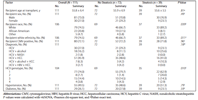
Table 1. Recipient Demographics and Pretransplant Characteristics
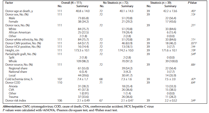
Table 2. Donor Characteristics
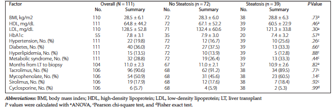
Table 3. Other Posttransplant Characteristics at 12 Months
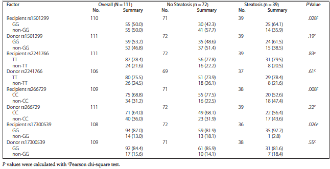
Table 4. Recipient and Donor Genotypes
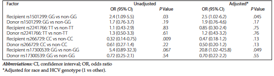
Table 5. Adjusted Comparison of Genotypes