Volume: 16 Issue: 4 August 2018
FULL TEXT
The number of kidney transplant candidates is increasing sharply. Among them, at least 20% are HLA sensitized. For these patients, in the setting of both living- and deceased-donor kidney transplant, we may face donor-specific alloantibodies at pretransplant. In such cases, the microlymphocytotoxicity crossmatch may or may not be positive. Kidney transplant with donor-specific antibodies at pretransplant is known as HLA-incompatible transplant. At present, we can use many methods to ensure that the kidney transplant is successful, provided that the recipient is desensitized before or after transplant. In this paper, we review the various strategies available for desensitization of HLA-incompatible kidney transplant recipients and the long-term outcomes.
Key words : Double-filtration plasmapheresis, IdeS, Immunoadsorption, Plasmapheresis, Renal transplantation, Rituximab, Tocilizumab
Introduction
The number of patients worldwide affected by end-stage renal disease is increasing sharply, in both developed and developing countries.1 Among patients with end-stage renal disease, many are suitable for kidney transplant. Compared with dialysis, kidney transplant is associated with significantly better quality of life2 and longer survival.3 Moreover, preemptive kidney transplant (ie, transplant before starting dialysis therapy) has been shown to be the best option because it is associated with significantly better long-term patient and graft survival rates than shown in patients who receive dialysis before transplant.4,5 Recipients can receive a living-donor (the best option) or a deceased-donor kidney transplant. In addition, recipients may receive multiple kidney transplants. However, each transplant will result in HLA sensitization, that is, having antibodies against HLA antigens of class I and/or II and making it difficult to find a subsequent HLA compatible transplant, that is, a donor against which the recipient has no donor-specific alloantibodies (DSAs) regardless of the number of HLA matches.
There are donor exchange programs in some countries that include both deceased and living kidney donors; therefore, for some HLA-sensitized kidney transplant candidates who have many anti-HLA antibodies, we may avoid the situation where a recipient has at transplant a DSA.6,7
In some settings, we transplant a kidney when the recipient has one or more DSAs. These transplant procedures are termed HLA incompatible. Nonetheless, it is of utmost importance to use biologic tests that are sensitive enough to detect DSAs. One method of detection is a microlymphocytotoxicity test. For this, only DSAs that can fix/activate complements are detected. However, we know that complement-activating DSAs are the most harmful as they result in a positive T-cell crossmatch. If present at the time of transplant, hyperacute rejection will occur. Fortunately, this situation now never occurs because complement-dependent cytotoxicity crossmatches (CDCs) are assessed immediately before transplant. Other DSAs do not fix complement and are detected using flow cytometry, enzyme-linked immunosorbent assay, or Luminex (Luminex, Austin, TX, USA).
At present, the most popular test, despite its expense, is Luminex, which enables detection of immunoglobulin G (IgG) antibodies directed against class I or class II HLA antigens using LABScreen mixed assays (One Lambda, Canoga Park, CA, USA). The results are reported as a normalized background ratio. When screened serum is positive, its reactivity is then assessed using a single-antigen bead assay (HLA class I and II LABScreen). The results are expressed as mean fluorescence intensity (MFI), with the cutoff point varying from one laboratory to another but generally between 500 and 1000 MFI. The higher the MFI value, the higher the likelihood of a positive crossmatch and potential to fix complement (ie, the C1q-binding or C3d-binding tests are positive).8-10 Thus, it is obvious that the higher the MFI value the higher the immunologic risk and the higher the difficulty in removing the DSA. Hence, having a DSA at pretransplant increases the risk of antibody-mediated rejection (AMR), increases the risk of transplant glomerulopathy, and increases the risk of allograft loss.11
The natural history of AMR, related to a preformed DSA or to a de novo DSA, can be expressed in 2 ways. In one setting, there will be indolent AMR; that is, DSAs will damage transplant endothelial cells, with subsequently complement activation and C4d deposits, resulting in peritubular capillaritis and glomerulitis. This persistent microvascular inflammation will lead to allograft dysfunction. In the second setting, DSAs will cause acute AMR, that is, acute renal failure with histologic patterns of glomerulitis and peritubular capillaritis. At this point, the patient receives a specific treatment for AMR, although not sufficient enough to avoid persistent microvascular inflammation, ultimately resulting in allograft dysfunction. When allograft dysfunction occurs, this translates into chronic AMR, ie, pathologic patterns of interstitial fibrosis/tubular atrophy, graft arteriosclerosis, and transplant glomerulopathy. At this stage, there is as yet no established efficient therapy to stop the process that leads to allograft loss.12 To avoid chronic AMR, overt and indolent AMR must be prevented. However, can this be achieved by desensitization? In this paper, we review the various strategies available for desensitization of HLA-incompatible kidney transplant recipients and the long-term outcomes (Table 1).
Defining Desensitization
Desensitization is usually applied at before transplant, although in some cases
after transplant to remove DSAs or to at least decrease their strength.13,14
Desensitization relies on removing the culprit DSA(s), which can be achieved by
apheresis, and preventing DSA resynthesis by targeting both T and B cells. It is
best to eliminate DSAs altogether; however, what happens if the DSA persists at
(very) low levels? Mohan and associates performed a meta-analysis to evaluate
the impact of low DSA levels at pretransplant, even if CDC and flow cytometry
crossmatches were negative. They found that the risk of AMR was then increased
by 98% and the risk of allograft loss was increased by 76%.15 In another study,
a Baltimore group showed that HLA-incompatible desensitized recipients who
received kidneys from living donors had significantly better long-term survival
than those on a kidney transplant wait list and who did not receive a kidney
from a deceased donor or those who were receiving dialysis therapy and not
suitable to be on a kidney transplant wait list.16
More recently, Orandi and associates reported the long-term results of > 1000 HLA-incompatible desensitized patients who received living-donor kidney transplant. The authors confirmed the results from the Baltimore group (ie, desensitized patients had better long-term survival than the other 2 above-mentioned categories).17 These multicenter results indicated that it is worth to desensitize the HLA-incompatible patient who has a potential living donor.18 However, we have few long-term data on the renal functionality of these desensitized patients.
The Mayo Clinic group compared 5-year data on HLA-incompatible desensitized living-donor kidney transplants versus HLA-compatible living-donor kidney transplants.19 They reported that graft survival was significantly lower in those with a positive crossmatch than in those with a negative crossmatch; however, graft survival was worse in those with a CDC-positive crossmatch compared with those with a flow cytometry crossmatch-positive crossmatch. They also found that those with a DSA against class II antigens had significantly worse outcomes than those who had a DSA against class I antigens.
Best Tools and Practices to Desensitize HLA-Incompatible Kidney Transplant
Recipients
If there is a living donor, desensitization takes place before transplant;
conversely, if there is a deceased donor, then desensitization takes place
immediately before transplant and is continued after transplant. When a
potential living donor is identified, clinicians can target high-risk groups,
that is, those with a positive T-cell crossmatch (using CDC). Conversely, if the
kidney is from a deceased donor and there are only a few hours available at
pretransplant, clinicians mostly target patients with a positive T-cell flow
cytometry crossmatch.
Tools before desensitization
Before a desensitization program is started, we need to know the MFI of the
potential DSA: the higher the MFI, the more complex is the required apheresis.
When the MFI of the DSA is < 3000 before transplant, apheresis is not necessary because the DSA strength is low. In these cases, we have to prevent the DSA from rebounding posttransplant. Typically, these patients are at high risk of an acute humoral rejection after transplant. The induction therapy could be based on rituximab and antithymocyte globulin (ATG), and maintenance immunosuppression is conventional (ie, tacrolimus, mycophenolic acid, and steroids). The posttransplant follow-up of DSAs has been already summarized in a consensus paper.20
When the MFI of the DSA is ≥ 3000 before transplant, apheresis should be attempted. The MFI could even be as high as 20 000. However, before desensitization is attempted, it is always mandatory to eliminate a prozone effect caused by competition between the fluorescent anti-IgG conjugate and serum complement for the alloantibody. This could be done using various methods, such as treating the serum with EDTA, dithiothreitol, heat inactivation, or serum dilution.8 Apheresis should always be implemented while the patient is already receiving immunosuppressive drugs.
Plasmapheresis is easy to set up but only permits treatment within 1 session of small volumes of plasma. The plasma removed must be replaced by either fresh frozen plasma or albumin. In developed countries, fresh frozen plasma is expensive. After each session of plasmapheresis, immunoglobulin rebound occurs (ie, interstitial immunoglobulins refill the vascular compartment). Thus, the use of plasmapheresis should be limited to cases where the highest MFI (of the DSAs) is ≤ 9000. In such cases, plasmapheresis must be performed on a daily basis, and its efficacy (ie, when the MFI becomes ≤ 3000) must be assessed after every 5 plasmapheresis sessions. The number of subsequent plasmapheresis sessions should be adapted accordingly. In the event of a DSA not reducing to < 3000 MFI, other apheresis techniques should be implemented.
If the MFI of the DSA is > 9000 but below 13 000, double-filtration plasmapheresis (DFPP) can be implemented. This process allows larger volumes of plasma to be treated compared with plasmapheresis; in addition, because pore sizes of the filters can be varied, large amounts of immunoglobulins can be cleared. However, the price for immunoglobulin clearance is that it also depletes large-molecular-weight clotting factors, such as fibrinogen, factor XIII, and von Willebrand factor. Although DFPP can be implemented on a daily basis, MFI must be assessed after every 5 sessions. In some cases, the frequency of DFPP sessions is limited by fibrinogen level, for example, when the fibrinogen level before a DFPP session is < 1 g/L. This process can be used as a starter therapy, that is, to decrease a DSA’s MFI to < 9000, and, when this target is reached, DFPP can be converted to plasmapheresis.
In the event that a DSA’s MFI is > 12 000 before starting desensitization, DFPP is insufficient; in these cases, semi-specific immunoadsorption has to be used. There are 2 types of columns on the market. One column contains highly purified protein A (isolated from Staphylococcus aureus) bound to a silica matrix (Immunosorba, Fresenius Medical Care, Bad Homburg, Germany). The other column, the Globaffin (Fresenius Medical Care) column, is a synthetic broadband immunoadsorber that contains a synthetic peptide (GAM) as a ligand. These 2 columns work in tandem; that is, when the plasma is running through one, regeneration takes place in the other. Both columns have a high affinity with a constant (Fc) segment, especially for IgG class antibodies. These 2 types of columns can be reused 10 to 20 times.
To enhance the efficacy of immunoadsorption columns, we can couple them with a membrane filter, such as a MONET (membrane filtration optimized novel extracorporeal treatment; Fresenius Medical Care), which enables the removal of large-molecular-weight molecules (ie, those with a molecular weight of > 1 kDa). Although semi-specific immunoadsorption does not affect the clotting factors, membrane filtration with MONET does.21 Therefore, MONET can be coupled with semi-specific immunoadsorption on an alternate-day basis. When the MFI is > 12 000, semi-specific immunoadsorption is conducted on a daily basis with MFI assessed once weekly to plan the next sessions. When the MFI is reduced (ie, < 6000), semi-specific immunoadsorption can be replaced by DFPP or plasmapheresis (see above) to obtain a MFI threshold of ~3000.
In all of these scenarios, as soon as MFI is reduced to < 3000, kidney transplant can be performed with ATG as the induction therapy. Regarding other immunosuppressive drugs, tacrolimus, mycophenolic acid, and steroids can be started as soon as the apheresis sessions have started. The question is whether other immunomodulatory drugs are required when starting apheresis.
Intravenous immunoglobulins
So far, intravenous immunoglobulins (IVIg) have only been tested in 2 randomized
clinical trials in order to desensitize kidney transplant candidates. One study
compared IVIg versus placebo and the other compared IVIg plus rituximab versus
IVIg plus a placebo. The first trial (NHI IG02) was conducted before Luminex
technology was available and when sensitization was expressed as a percentage of
panel-reactive alloantibodies (PRA). Thus, interpretation of these results
should be cautionary. Study patients had to have PRA > 50%.22 Of 98 trial
patients, 48 were randomized to the IVIg arm and received IVIg at
2 g/kg once per month, scheduled for 4 months, and then 2 g/kg at months 12 and
24. The follow-up was 30 months. Intravenous immunoglobulin significantly
reduced PRA levels compared with placebo and significantly reduced time until
transplant. Sixteen IVIg patients (35%) and 8 of the 50 placebo patients (17%)
received transplants. However, 59% of IVIg patients and 10% of placebo patients
(P = .04) experienced acute rejection. The 2-year patient and graft survival
rates were similar across the 2 groups.
Conversely, Alachkar and associates enrolled 27 highly sensitized kidney transplant candidates (calculated PRA = 100%) who were given IVIg at 2 g/kg monthly for 4 months: these authors reported that IVIg did not affect the calculated PRA.23
Vo and associates recently reported on a randomized double-blind control trial that included highly sensitized kidney transplant candidates and compared IVIg plus a placebo with IVIg plus rituximab. The trial was stopped after 15 patients were enrolled and was unblinded because 5 serious adverse events occurred. Of the 15 patients, 13 received a transplant. All episodes of acute AMR occurred in the IVIg plus placebo arm and required intense therapy (P = .06). The 2 graft losses were also in the placebo group. DSA rebound was only seen in the placebo group and was associated with severe acute AMR; conversely, no DSA rebound was reported in the IVIg plus rituximab group.24 Thus, these trials show that IVIg alone was unable to desensitize kidney transplant candidates.
Intravenous immunoglobulins plus rituximab, with or without apheresis
Only 1 randomized trial has been conducted on IVIg plus rituximab (see above).
However, the largest single-center study is from Cedars Sinai Hospital (Los
Angeles, CA, USA), which included 207 sensitized patients in which
desensitization relied on IVIg (2 g/kg twice, at 1 month apart) plus rituximab
(initially 500 mg twice and thereafter a 1-g single injection).25 Induction
therapy included alemtuzumab, ATG, or daclizumab. Maintenance immunosuppression
included tacrolimus, mycophenolate mofetil (MMF), and steroids. Desensitization
treatment was associated with significantly decreased PRA level
and T-cell flow cytometry crossmatch, as assessed
by decreased mean channel shift. Of the 207 desensitized patients, 146 (70.5%)
received a kidney transplant. At 48 months follow-up, patient and graft survival
rates were 95% and 87.5%, respectively. For those who received a transplant,
independent predictors for AMR were having had a previous transplant, the
relative intensity of DSA, the presence of both class I and class II DSAs at
transplant, and time on the wait list. Patients who had AMR had significantly
reduced graft survival and significantly reduced glomerular filtration rates at
5 years, especially those who have had thrombotic microangiopathy.
Magee and associates reported their experience on plasmapheresis, IVIg, and rituximab given to 28 renal transplant candidates who had a potential living donor with a positive lymphocytotoxic crossmatch (T-cell-positive or B-cell-positive). Eleven patients had AMR; within a mean follow-up of 22 months, only 3 grafts were lost, and mean serum creatinine level was good, at 1.5 mg/dL.26
Plasmapheresis, intravenous immunoglobulins, and rituximab
In the setting of deceased-donor kidney transplant, the Necker Hospital group
(Paris) reported on a series of patients who received a transplant in the
setting of a DSA on the day of transplant but who had negative T-cell
crossmatches. In this situation, the historical cohort (n = 36) was desensitized
at posttransplant with IVIg 2 g/kg at days 0, 2, 42, and 63 or were part of a
cohort (n = 18) who also received rituximab at 375 mg/mL on day 2 and day 22
plus 9 plasmapheresis sessions on an alternate-day basis at posttransplant. All
patients underwent a kidney biopsy on day 90 and day 365. At 1-year
posttransplant, patient and graft survival rates were similar between the 2
groups as was the rate of acute AMR. Conversely, estimated glomerular filtration
rate was significantly worse and proteinuria
was significantly higher in the IVIg group, as
well as the rate of chronic AMR (41.3% vs 13.3%;
P = .03). Moreover, the decline in DSA’s MFI from day 0 to 1 year posttransplant
was 44% ± 13% in the IVIg group compared with 80% ± 8% in the
plasmapheresis/rituximab/IVIG group (P = .02).27
Recently, the same group reported the long-term results of 95 sensitized
patients who received a transplant from a deceased donor against which they had
at least one DSA with a MFI of > 3000 and a negative CDC crossmatch.
Desensitization relied on plasmapheresis, IVIg, and rituximab. The authors
compared these results versus those from 39 patients who had DSAs against the
deceased donor (MFI of
< 3000) and in which posttransplant desensitization was based on IVIg alone.
Patient survival was the same; however, graft survival was significantly lower
for those with MFI > 3000 (allograft loss 28.4% vs 5.13%; P = .002). In
addition, there were significantly more cases of acute T-cell rejection (11.6%
vs 0%) and AMR (32.6% vs 10.3%; P = .005) in the group with MFI > 3000. The
authors concluded that their desensitization strategy was worth it, even when
the DSA MFI was > 3000, with a deceased donor.28
Immunoadsorption plus rituximab
Semi-specific immunoadsorption can use Immunsorba or Globaffin tandem columns
(see above). The pioneer of this strategy was a group from Vienna, Austria.29
The group recently reported on a series of 101 DSA-positive recipients of
deceased-donor kidney transplant and who had been desensitized since 2009;
median posttransplant follow-up was 24 months. Before transplant, 27 of the 101
recipients had a positive CDC crossmatch. All patients received an
immunoadsorption session before transplant (for those who had a CDC-positive
crossmatch before, this had to become negative after the session).
Immunoadsorption was performed on a daily basis postoperatively to decrease MFI
to < 3000. All patients were induced with ATG; maintenance immunosuppression
relied on tacrolimus, MMF, and steroids. The authors compared these results with
those from a control group consisting of 513
DSA-negative kidney transplant recipients treated during the same period. The
3-year graft-survival rate in DSA-positive patients was 79% compared with 88% in
DSA-negative patients (P = .008). One-third of DSA-positive patients experienced
acute AMR; their DSA MFI value was significantly associated with acute rejection
(ie, 20% vs 71% AMR with a MFI of < 5000 compared with > 15 000 peak MFI,
respectively). The predictive value of the MFI
was moderate. The authors concluded that immunoadsorption-based desensitization
failed to prevent AMR in one-third of DSA-positive patients in the setting of
deceased-donor kidney transplant and that assessing the MFI could help stratify
for risk of rejection. This could be used as a guide during organ allocation.30
Kauke and associates recently reported on a small series of 8 Luminex-assessed patients with positive DSAs who were desensitized with immunoadsorption and rituximab in the setting of living-donor kidney transplant. One-year graft survival rates and renal function (1.78 ± 0.6 mg/dL) were excellent; the incidence of T-cell-mediated rejection was 12.5%, whereas that of acute AMR was 21%.31
A group in Heidelberg reported on a series of 23 desensitized DSA-positive recipients of living-donor kidney transplant where the goal was to maintain MFI at < 1000. Patients had an average of 8 (range, 1-23) immunoadsorption sessions before transplant plus received tacrolimus, MMF, steroids, and 1 rituximab injection during days 0 to 1. The induction therapy was based on either ATG or basiliximab. Immunoadsorption sessions were maintained immediately after transplant, resulting in an average of 5 sessions (range, 0-18) until serum creatinine became < 2 mg/dL and MFI was < 1000. These results showed that, at day 0, median MFI was < 1000. The 2-year graft survival rate was 100%, with excellent renal function (median serum creatinine level of 1.42 mg/dL) and protein-to-creatinine ratio of 0.12 g/L. Only 9% of patients had a persistent DSA, and only 9% had an emerging DSA. Finally, 21% had an AMR; these results are encouraging.32
Rostaing and associates reported on 7 highly sensitized patients who were DSA-positive against their living kidney donor.33 The patients were desensitized by IVIg, rituximab, and immunoadsorption (10-16 sessions/patient); all were induced with ATG. At transplant, DSAs were either undetectable or MFI levels had decreased by > 50% in 31% of the patients; 3 patients presented with an AMR. Long-term renal functionality was good.
Other strategies
In the United States, the only apheresis technique that is FDA approved is
plasmapheresis. The largest series of HLA desensitization based on
plasmapheresis plus low immunoglobulin (anti-cytomegalovirus immunoglobulin at
100 mg/kg after each plasmapheresis session) was reported by Montgomery and
associates.16 All transplant candidates received a living kidney transplant and
had DSAs, with or without a positive crossmatch (CDC or flow cytometry).
Immunosuppression included ATG or basiliximab as induction therapy, plus
maintenance immunosuppression based on tacrolimus, MMF, and steroids. On
average, patients received 4 ± 4 plasmapheresis sessions at before transplant
and 5 ± 4 plasmapheresis sessions after transplant (number of sessions was
higher pre- and posttransplant for those who had a CDC-positive crossmatch). The
authors found that survival of patients at 5 years was significantly greater for
sensitized patients (90.6%) than in wait list patients who remained on dialysis
(51.5%) or in those placed on a deceased-donor kidney transplant wait list with
or without kidney transplant (65.6%).
From this series, between 2000 and 2010, 129 HLA-incompatible patients received a transplant with a positive crossmatch (follow-up of 1-9 y), in which 745 follow-up kidney allograft biopsies were conducted.34 At 5 years, allograft survival was 80%. Throughout the follow-up, 70% of patients had at least 1 rejection episode with 52% presenting with a subclinical rejection, seen on the 1-year surveillance biopsy. In addition, throughout follow-up, cellular rejection was more frequent than AMR. In addition, transplant glomerulopathy developed in 47% of patients over the period of the study; transplant glomerulopathy was preceded by glomerulitis. Significant risk factors for transplant glomerulopathy included glomerulitis and a detectable DSA at posttransplant. Finally, C4d-negative AMR, manifesting as capillaritis (glomeruli ≥ 1 and peritubular capillaries ≥ 1) with a detectable DSA, was observed in 20% of the recipients. It was concluded that, despite good graft survival, these HLA-incompatible, desensitized, living-donor kidney recipients needed to be closely monitored due to the subclinical rejection.34
The long-term survival benefit of patients who had been desensitized and were recipients of a living donor kidney reported by Montgomery and associates16 was confirmed in a larger multicenter (n = 22) US study that included 1025 patients. At 5-year follow-up, overall patient survival was 86% compared with only 59.2% for patients receiving dialysis and 74.4% for patients on wait list after receiving or not a deceased-donor kidney transplant during follow-up (P < .001). This was also the case in a subgroup of patients who had positive T-cell crossmatches before being started on desensitization. However, we do not have functional data and do not know how the patients were desensitized.17
Other Therapies, Including Monoclonal Antibodies That Interfere With the Complement Pathway
Tocilizumab
Tocilizumab is a monoclonal antibody directed against the interleukin 6
receptor. It blocks the activity of interleukin 6 and may interfere with
plasmablasts/plasma cells, thereby decreasing the production of DSAs. No
randomized control trial has been conducted in the setting of desensitization.
In a single-center study (from Cedars Sinai Hospital), 10 patients who were
unresponsive to IVIg plus rituximab were then given tocilizumab at 8 mg/kg
monthly for up to 6 months. If they were to receive a transplant, tocilizumab
was continued on a monthly basis at posttransplant. Of the 10 patients, 5
received a transplant.35 All treated patients had a decreased MFI of the
immunodominant DSA, and this decrease was prolonged and increased within the
first year for transplant recipients. More data on this treatment are needed.
Eculizumab
Eculizumab is a monoclonal antibody that blocks
the C5a. In a desensitization study from the Mayo
Clinic (USA), living-donor kidney recipients with pretransplant DSAs but
negative crossmatches were given eculizumab injections (at days 0 and 1, then
weekly for 1 month, thereafter every 2 weeks) for 1 year posttransplant.36 In
addition, patients received ATG, tacrolimus, MMF, and steroids (n = 26). The
control historical group (n = 51) consisted of living-donor kidney recipients
with pretransplant DSAs who had received pre- and posttransplant plasmapheresis
plus tacrolimus, MMF, and steroids. At 1-year posttransplant, patient and graft
survival rates were similar between the groups, although there were
significantly more cases of AMR in the control group (41% vs. 7.7%; P = .03) and
transplant glomerulopathy shown in the 1-year protocol biopsies (36% vs. 6.7%;
P
= .04).36 Since this paper was published, Alexion (Cheshire, CT, USA) has
conducted a phase II trial in which 102 patients received a kidney transplant
from a living donor, and all recipients were at risk for AMR, based on elevated
DSA levels.37 The primary composite endpoint was the occurrence of biopsy-proven
AMR, graft loss, patient death, or lost to follow-up at week 9 posttransplant.
Consented patients were randomized into 2 groups of 51 patients each, with one
group receiving eculizumab and a control group receiving the anti-rejection
standard-of-care specified by the institution in which each patient’s
transplantation took place. In both groups, immunosuppression included
tacrolimus, MMF, steroids, and ATG as induction therapy. Patients in the control
arm were eligible to receive eculizumab treatment for presumed AMR. At 9 weeks,
analyses showed that 9.8% of patients in the eculizumab arm and 15.7% of
patients in the control arm (P = .554) had met the primary composite endpoint.
No safety signal was reported by the independent data monitoring committee.
Although the primary composite endpoint for the eculizumab arm was as expected
(from earlier studies with eculizumab), control arm numbers were lower than
expected, based on natural history studies reported in the literature.37
C1 esterase inhibitor
The C1-INH has been tested in highly sensitized renal transplant recipients to
prevent AMR. In one study, 20 highly sensitized patients who were desensitized
with IVIg plus rituximab with or without plasma exchange were randomized 1:1 to
receive plasma-derived human C1-INH (20 IU/kg/dose) versus a placebo
intraoperatively, which was then given twice weekly for 7 doses.38 One patient
in the C1-INH group versus 2 patients in the placebo group developed serious
adverse events; however, none were related to drugs used in the study. Delayed
graft function developed in 1 patient in the C1-INH group and in 4 patients in
the placebo control
group. The C1-INH trough levels increased with
C1-INH treatment. C3 and C4 levels also increased significantly in the C1-INH
group compared with the placebo group. No C1-INH patient developed AMR during
the study, although 2 patients developed AMR after the study. Three patients
from the placebo group developed AMR (1 during the study). C1q-positive DSAs
became reduced in 2 C1-INH-treated patients, whereas IgG DSA levels decreased
binding in both groups. The authors concluded that C1-INH appeared to be safe in
the posttransplant period. Treatment with C1-INH may reduce ischemia-reperfusion
injury, and treatment also resulted in significant elevations of C1-INH, C3, and
C4 levels and reduced C1q-positive HLA antibodies. Together, the combination of
antibody reduction and C1-INH may prove useful to prevent AMR.38
Bortezomib
There are few studies in humans regarding desensitization and bortezomib, and
results are inconclusive. Kwun and associates recently demonstrated, in a
primate model, that bortezomib induced humoral compensation in allo-sensitized
recipients. It was administered intravenously at 1.3 mg/m2 twice weekly for 1
month. They found that bortezomib
did not modify DSA levels and was associated with a rapid but transient
induction of circulating
IgG-positive B cells and an increased number of proliferating B cells in the
lymph nodes after 1 month of treatment. In addition, bortezomib induced germinal
center B-cell and follicular helper T-cell expansion in the lymph nodes.39
In a recent report, a Mayo clinic group40 aimed to determine the safety and efficacy of 32 doses of bortezomib (1.3 mg/m2) given to 10 highly sensitized kidney transplant candidates with alloantibodies against their intended living donor. Dose reduction was needed for 2 patients and 2 others discontinued therapy for adverse events. Anti-HLA antibody MFI values were stable before bortezomib treatment (P = .96) but decreased after therapy (mean [standard deviation] decrease of 1916 MFI [425 MFI]; P < .01). No patient developed a negative crossmatch against their original intended donor, and the calculated PRAs, based on MFIs of 2000, 4000, and 8000, were unchanged in all patients. The authors concluded that 32 doses of bortezomib (as a monotherapy) was not well tolerated and resulted in only a modest reduction in anti-HLA antibodies.40
A Korean group reported similar results; that is, bortezomib had a marginal impact on decreasing DSAs.41 This was a prospective, open-labeled clinical trial, in which 19 highly sensitized kidney transplant candidates, without a potential living kidney donor, were desensitized. The desensitization regimen consisted of 2 doses of IVIg (2 g/kg), a single dose of rituximab (375 mg/m2), and 4 doses of bortezomib (1.3 mg/m2). Anti-HLA antibodies were determined at baseline and after 2, 3, and 6 months in the desensitized patients. These 19 patients were compared with 17 matched highly sensitized patients who had not undergone desensitization therapy. Baseline values of class I and II PRAs were 83% ± 16.0% (14 952 ± 5820 peak MFI) and 63% ± 36.0% (10 321 ± 7421 peak MFI), respectively. Deceased donor-kidney transplant was successfully performed in 8 patients (42.1%) in the desensitization group versus 4 (23.5%) in the control group. Multivariate time-varying covariate Cox regression analyses showed that desensitization increased the probability of deceased-donor kidney transplant (hazard ratio of 46.895; 95% confidence interval, 3.468-634.132; P = .004). However, this desensitization protocol did not affect class I or II PRAs; it only significantly decreased peak class I PRA MFI values by 15.5% (20.8%) at 2 months (P = .002).41
Endoglycosidase and immunoglobulin G-degrading enzyme of Streptococcus pyogenes
Endoglycosidase (EndoS) and the IgG-degrading enzyme of Streptococcus pyogenes
(IdeS) are immunomodulating enzymes that hydrolyze human IgG: IdeS cleaves IgG
in the lower hinge region, whereas EndoS hydrolyzes the conserved N-linked
glycan in the Fc region. Both enzymes are remarkably specific to human IgG,
which, after hydrolysis, loses most of its effector functions, such as binding
to leukocytes and complement activation, which contribute to bacterial evasion
of adaptive immunity. It has been shown that IdeS and EndoS can alleviate
autoimmune disease in a number of animal models of antibody-mediated disorders.
There is a rationale for the development of IdeS and EndoS against diseases
where IgG antibodies contribute to the pathology (eg, chronic and acute
autoimmunity, transplant rejection, and antidrug antibody reactions).42 At
present, a phase I/II trial is being conducted that includes kidney transplant
recipients and is assessing the prevention of AMR in the setting of
pretransplant DSAs. The results are encouraging.43
Conclusions
Although evidence-based data are scarce in the field of HLA desensitization, some data have shown the feasibility of HLA-incompatible kidney transplant (where the recipient has 1 or more DSAs against the potential donor) and have shown that good long-term results can be achieved. The best strategy is based on apheresis to decrease MFI to < 3000 plus the use of rituximab. The addition of IVIg might also be valuable. In the near future, the perioperative use of IdeS could be promising because it may eliminate the risk of AMR, at least for the first 2 weeks posttransplant.
References:
- . Robinson BM, Akizawa T, Jager KJ, Kerr PG, Saran R, Pisoni RL. Factors affecting outcomes in patients reaching end-stage kidney disease worldwide: differences in access to renal replacement therapy, modality use, and haemodialysis practices. Lancet. 2016;388(10041):294-306.
CrossRef - PubMed - Niu SF, Li IC. Quality of life of patients having renal replacement therapy. J Adv Nurs. 2005;51(1):15-21.
CrossRef - PubMed - Ingsathit A, Kamanamool N, Thakkinstian A, Sumethkul V. Survival advantage of kidney transplantation over dialysis in patients with hepatitis C: a systematic review and meta-analysis. Transplantation. 2013;95(7):943-948.
CrossRef - PubMed - Goldfarb-Rumyantzev A, Hurdle JF, Scandling J, et al. Duration of end-stage renal disease and kidney transplant outcome. Nephrol Dial Transplant. 2005;20(1):167-175.
CrossRef - PubMed - Huang Y, Samaniego M. Preemptive kidney transplantation: has it come of age? Nephrol Ther. 2012;8(6):428-432.
CrossRef - PubMed - Huh KH, Kim MS, Ju MK, et al. Exchange living-donor kidney transplantation: merits and limitations. Transplantation. 2008;86(3):430-435.
CrossRef - PubMed - Fumo DE, Kapoor V, Reece LJ, et al. Historical matching strategies in kidney paired donation: the 7-year evolution of a web-based virtual matching system. Am J Transplant. 2015;15(10):2646-2654.
CrossRef - PubMed - Eskandary F, Bond G, Kozakowski N, et al. Diagnostic contribution of donor-specific antibody characteristics to uncover late silent antibody-mediated rejection-results of a cross-sectional screening study. Transplantation. 2017;101(3):631-641.
CrossRef - PubMed - Arreola-Guerra JM, Castelan N, de Santiago A, et al. C1Q Assay results in complement-dependent cytotoxicity crossmatch negative renal transplant candidates with donor-specific antibodies: high specificity but low sensitivity when predicting flow crossmatch. J Transplant. 2016;2016:2106028.
CrossRef - PubMed - Viglietti D, Loupy A, Vernerey D, et al. Value of donor-specific anti-HLA antibody monitoring and characterization for risk stratification of kidney allograft loss. J Am Soc Nephrol. 2017;28(2):702-715.
CrossRef - PubMed - Morath C, Opelz G, Zeier M, Susal C. Clinical relevance of HLA antibody monitoring after kidney transplantation. J Immunol Res. 2014;2014:845040.
CrossRef - PubMed - Loupy A, Hill GS, Jordan SC. The impact of donor-specific anti-HLA antibodies on late kidney allograft failure. Nat Rev Nephrol. 2012;8(6):348-357.
CrossRef - PubMed - Wongsaroj P, Kahwaji J, Vo A, Jordan SC. Modern approaches to incompatible kidney transplantation. World J Nephrol. 2015;4(3):354-362.
CrossRef - PubMed - Jordan SC, Choi J, Vo A. Kidney transplantation in highly sensitized patients. Br Med Bull. 2015;114(1):113-125.
CrossRef - PubMed - Mohan S, Palanisamy A, Tsapepas D, et al. Donor-specific antibodies adversely affect kidney allograft outcomes. J Am Soc Nephrol. 2012;23(12):2061-2071.
CrossRef - PubMed - Montgomery RA, Lonze BE, King KE, et al. Desensitization in HLA-incompatible kidney recipients and survival. N Engl J Med. 2011;365(4):318-326.
CrossRef - PubMed - Orandi BJ, Luo X, Massie AB, et al. Survival benefit with kidney transplants from HLA-incompatible live donors. N Engl J Med. 2016;374(10):940-950.
CrossRef - PubMed - Rostaing LP, Malvezzi P. HLA-incompatible kidney transplantation--worth the risk? N Engl J Med. 2016;374(10):982-984.
CrossRef - PubMed - Bentall A, Cornell LD, Gloor JM, et al. Five-year outcomes in living donor kidney transplants with a positive crossmatch. Am J Transplant. 2013;13(1):76-85.
CrossRef - PubMed - Tait BD, Susal C, Gebel HM, et al. Consensus guidelines on the testing and clinical management issues associated with HLA and non-HLA antibodies in transplantation. Transplantation. 2013;95(1):19-47.
CrossRef - PubMed - Biesenbach P, Eskandary F, Ay C, et al. Effect of combined treatment with immunoadsorption and membrane filtration on plasma coagulation--Results of a randomized controlled crossover study. J Clin Apher. 2016;31(1):29-37.
CrossRef - PubMed - Jordan SC, Tyan D, Stablein D, et al. Evaluation of intravenous immunoglobulin as an agent to lower allosensitization and improve transplantation in highly sensitized adult patients with end-stage renal disease: report of the NIH IG02 trial. J Am Soc Nephrol. 2004;15(12):3256-3262.
CrossRef - PubMed - Alachkar N, Lonze BE, Zachary AA, et al. Infusion of high-dose intravenous immunoglobulin fails to lower the strength of human leukocyte antigen antibodies in highly sensitized patients. Transplantation. 2012;94(2):165-171.
CrossRef - PubMed - Vo AA, Choi J, Cisneros K, et al. Benefits of rituximab combined with intravenous immunoglobulin for desensitization in kidney transplant recipients. Transplantation. 2014;98(3):312-319.
CrossRef - PubMed - Vo AA, Petrozzino J, Yeung K, et al. Efficacy, outcomes, and cost-effectiveness of desensitization using IVIG and rituximab. Transplantation. 2013;95(6):852-858.
CrossRef - PubMed - Magee CC, Felgueiras J, Tinckam K, Malek S, Mah H, Tullius S. Renal transplantation in patients with positive lymphocytotoxicity crossmatches: one center's experience. Transplantation. 2008;86(1):96-103.
CrossRef - PubMed - Loupy A, Suberbielle-Boissel C, Zuber J, et al. Combined posttransplant prophylactic IVIg/anti-CD 20/plasmapheresis in kidney recipients with preformed donor-specific antibodies: a pilot study. Transplantation. 2010;89(11):1403-1410.
CrossRef - PubMed - Amrouche L, Aubert O, Suberbielle C, et al. Long-term outcomes of kidney transplantation in patients with high levels of preformed DSA: the Necker high-risk transplant program. Transplantation. 2017;101(10):2440-2448.
CrossRef - PubMed - Lorenz M, Regele H, Schillinger M, et al. Peritransplant immunoadsorption: a strategy enabling transplantation in highly sensitized crossmatch-positive cadaveric kidney allograft recipients. Transplantation. 2005;79(6):696-701.
CrossRef - PubMed - Schwaiger E, Eskandary F, Kozakowski N, et al. Deceased donor kidney transplantation across donor-specific antibody barriers: predictors of antibody-mediated rejection. Nephrol Dial Transplant. 2016;31(8):1342-1351.
CrossRef - PubMed - Kauke T, Klimaschewski S, Schoenermarck U, et al. Outcome after desensitization in HLA or ABO-incompatible kidney transplant recipients: a single center experience. PLoS One. 2016;11(1):e0146075.
CrossRef - PubMed - Klein K, Susal C, Schafer SM, et al. Living donor kidney transplantation in patients with donor-specific HLA antibodies enabled by anti-CD20 therapy and peritransplant apheresis. Atheroscler Suppl. 2013;14(1):199-202.
CrossRef - PubMed - Rostaing L, Congy N, Aarnink A, et al. Efficacy of immunoadsorption to reduce donor-specific alloantibodies in kidney-transplant candidates. Exp Clin Transplant. 2015;13 Suppl 1:201-206.
CrossRef - PubMed - Bagnasco SM, Zachary AA, Racusen LC, et al. Time course of pathologic changes in kidney allografts of positive crossmatch HLA-incompatible transplant recipients. Transplantation. 2014;97(4):440-445.
CrossRef - PubMed - Vo AA, Choi J, Kim I, et al. A Phase I/II trial of the interleukin-6 receptor-specific humanized monoclonal (tocilizumab) + intravenous immunoglobulin in difficult to desensitize patients. Transplantation. 2015;99(11):2356-2363.
CrossRef - PubMed - Stegall MD, Diwan T, Raghavaiah S, et al. Terminal complement inhibition decreases antibody-mediated rejection in sensitized renal transplant recipients. Am J Transplant. 2011;11(11):2405-2413.
CrossRef - PubMed - Alexion. Alexion Provides Update on Phase 2 Clinical Trial with Eculizumab in Antibody Mediated Rejection (AMR) in Living-Donor Kidney Transplant Recipients. Cheshire, CT: Alexion; January 7, 2015.
- Vo AA, Zeevi A, Choi J, et al. A phase I/II placebo-controlled trial of C1-inhibitor for prevention of antibody-mediated rejection in HLA sensitized patients. Transplantation. 2015;99(2):299-308.
CrossRef - PubMed - Kwun J, Burghuber C, Manook M, et al. Humoral compensation after bortezomib treatment of allosensitized recipients. J Am Soc Nephrol. 2017;28(7):1991-1996.
CrossRef - PubMed - Moreno Gonzales MA, Gandhi MJ, Schinstock CA, et al. 32 Doses of bortezomib for desensitization is not well tolerated and is associated with only modest reductions in anti-HLA antibody. Transplantation. 2017;101(6):1222-1227.
CrossRef - PubMed - Jeong JC, Jambaldorj E, Kwon HY, et al. Desensitization using bortezomib and high-dose immunoglobulin increases rate of deceased donor kidney transplantation. Medicine (Baltimore). 2016;95(5):e2635.
CrossRef - PubMed - Collin M, Bjorck L. Toward clinical use of the IgG specific enzymes IdeS and EndoS against antibody-mediated diseases. Methods Mol Biol. 2017;1535:339-351.
CrossRef - PubMed - Jordan SC, Lorant T, Choi J, et al. IgG endopeptidase in highly sensitized patients undergoing transplantation. N Engl J Med. 2017;377(5):442-453.
CrossRef - PubMed

Volume : 16
Issue : 4
Pages : 367 - 375
DOI : 10.6002/ect.2017.0355
From the 1Service de Néphrologie, Hémodialyse, Aphérèses et Transplantation,
Centre Hospitalier Universitaire Grenoble-Alpes, Grenoble, France; the
2Université Joseph Fourier, Grenoble Alpes, France; and the 3INSERM U563,
IFR-BMT, Centre Hospitalier Universitaire Purpan, Toulouse, France
Acknowledgements: The authors have no sources of funding for this study and have
no conflicts of interest to declare. *Paolo Malvezzi and Thomas Jouve
contributed equally to this review.
Corresponding author: Lionel Rostaing, Service de Néphrologie, Hémodialyse,
Aphérèses et Transplantation, Centre Hospitalier Universitaire Grenoble-Alpes,
Avenue du maquis du Grésivaudan, 38700 La Tronche, France
Phone: +33 476768945
E-mail: lrostaing@chu-grenoble.fr
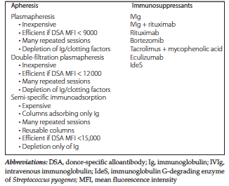
Table 1. Tools Available to Desensitize HLA-Incompatible Kidney Transplant Candidates